
DIAGNOSTIC
What is MRI?
Magnetic resonance imaging (MRI) is a diagnostic imaging technique based on the use of the phenomenon of nuclear magnetic resonance and has nothing to do with charged radioactive particles and other things potentially hazardous to health.MRI does not use penetrating radiation that is harmful to health. And that's why the task during the study is to create such conditions under which the patient's body itself will become a source of extremely weak radio signals. Such radio signals exist without any additional equipment and conditions, since this is one of the indicators of the correctness of the course of life processes in our body.
In MRI, we only isolate one patient - one source - from all other radio sources to minimize the effects of interference. Then, using the hardware of the tomography, the chaotic signals are ordered so that they can only be received from a specific area of the body. This requires a powerful but harmless to humans, magnetic field. Then, using very sensitive antennas and receivers, the radio signal is received, processed by an ultra-high-speed computer and an image is obtained. It reflects the distribution of radio signals of the cells of the human body in different planes and ratios. In medicine, such an image is called a tomogram. This is the most important step. Normal cells of organs and tissues not affected by the disease process have the same signal level. "Diseased" cells are always a different, altered signal to one degree or another.
On the image, the areas of tissues and organs changed by the pathological process look different than healthy ones. This is the basis of medical diagnostic imaging. Recognizing detailed changes is a matter of the experience of the medical researcher, a derivative of the quality of the apparatus. The main task is to obtain the most informative image quickly and efficiently, with comfort for the patient.


What we can?
What tasks are we solving? Perhaps, here it is necessary to focus on the main sections of our activity. The most complete information can be obtained by contacting us on a specific issue.Tomograms contain a huge amount of information about the structure of organs and tissues in a particular anatomical area. The structure, the relationship of organs to each other, their size and configuration - these are the main points that we evaluate in the course of the study. The magnetic resonance imaging scanner is designed in such a way that it is impossible to examine all areas of interest during one session. This means that one of the most important tasks for a doctor who sends his patient for an MRI is to accurately indicate to our doctor the anatomical area in which the study is to be performed. In addition, there is a wide variety of special programs and research methods. Each of these programs is prepared to identify the characteristic signs of a certain group of pathological changes. Therefore, conducting an MRI study for all programs at once is an extremely long and very expensive procedure.
Like other medical examinations, MRI is used to detect those diseases that cannot be detected in any other way. Or another method is unacceptable due to, for example contraindications. This means that the treating doctor who sends a patient for an MRI must formulate a specific goal of such a direction.
What is computed tomography?
Computed tomography is a diagnostic imaging technique that is much more informative than a conventional X-ray. The word "tomography" comes from the Greek words "tomos", which means layer or slice, and "graphy", which means to display.
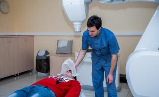
How is the study on MSCT carried out?
You are laid on a special table. While the table is moving, the selected area is scanned. The received data is displayed on the computer screen and stored in its memory. Modern software allows you to obtain a three-dimensional image of the organs of the body systems. You receive the results of the study, printed on a special printer, in a form familiar to you.
Let's note the advantages of MSCT over conventional computed tomography:
- dramatic improvement in image quality
- an increase in the scanning speed, and as a consequence, a decrease in the research time
- improved contrast resolution
- increase in signal-to-noise ratio
- large area of anatomical coverage
- reduction of radiation exposure to the patient
All these factors significantly increase the speed and information content of research.
As part of the study, you may be offered the introduction of a contrast agent. Contrast enhancement techniques allow you to determine and distinguish the nature of the identified changes, when this is not possible in a conventional study.
Our computed tomography center will provide your doctor with all the information he needs to make a correct diagnosis and choose the optimal treatment.
The high level of professionalism of doctors and medical technicians allows us to carry out studies of all organs and systems of the body of any complexity.
You'll get:
- a written opinion of the doctor based on the results of the study (usually within 24 hours),
- Disc with recording of images of your examination.
- Restrictions on conducting computed tomography studies
- intolerance to preparations containing iodine (for studies with contrast enhancement);
- the patient's condition, which does not allow holding his breath for a long time (more than 20 seconds);
- the body weight of the subject is more than 150 kg;
- the presence of barium suspension in the intestine;
- the presence of a plaster cast and (or) a metal structure in the research area;
- pronounced claustrophobia;
- Pregnancy and breastfeeding.
Positron emission tomography (PET)
Positron emission tomography (PET) is a modern diagnostic method that allows you to identify pathology by assessing biochemical changes in tissues. The method is based on the use of radiopharmaceuticals (RFP) in the minimum doses that are safe for the body. Depending on the activity of the functional process in various tissues of the body, the distribution of radiopharmaceuticals is displayed in the form of a color image of varying intensity.Positron emission tomography (PET) is a modern diagnostic method that allows you to identify pathology by assessing biochemical changes in tissues. The method is based on the use of radiopharmaceuticals (RFP) in the minimum doses that are safe for the body. Depending on the activity of the functional process in various tissues of the body, the distribution of radiopharmaceuticals is displayed in the form of a color image of varying intensity. To determine the exact localization of the foci of accumulation of RP, positron emission tomography is combined with computed tomography (CT). This combined PET / CT diagnostic method is used in oncology, neurology and cardiology.
The effectiveness of the method in oncology is due to the fact that PET / CT allows you to determine the stage of the pathological process, its prevalence and evaluate the effectiveness of the treatment.
The function of using PET / CT is extremely important when planning radiation therapy. Due to the fact that the method makes it possible to determine the tumor boundaries as accurately as possible and as well as the structural and functional characteristics of the affected and surrounding tissues, a clearer target for radiation exposure is formed, the risk of irradiation of healthy cells is significantly reduced and the side effects of radiation therapy are reduced.
In neurology, PET / CT is most often used to identify the localization of an epileptogenic focus, diagnose diseases associated with dementia (Alzheimer's disease, frontotemporal dementia, etc.), and assess the prevalence of post-traumatic and post-ischemic changes in brain tissue.
Studies of PET-CT with 18F-fluorodeoxyglucose and PET-CT with 18-FET fluoroethyltyrosine are carried out at the address: SPB, Glukharskaya str., Building 16, building 2 (entrance from Aviakonstruktorov street).
Timing of PET / CT to assess the effectiveness of treatment:
- 4-6 weeks after surgery.
- After 2 courses of PCT (if an assessment of the sensitivity of the tumor to the selected treatment regimen is required).
- 4-6 weeks after the last injection of PCT (if an assessment of the effectiveness of completed therapy is required).
- 2-4 weeks after radioiodine therapy.
- 6-8 weeks after the last radiation session.
Before the introduction of hormonal drugs in the last week.
PET / CT with 18F-FDG
Visit plan and preparation for the study
Among all radiopharmaceuticals, 18F-fluorodeoxyglucose is most often used, which is considered universal, since it is absorbed by almost all cells of the body because this RP is similar in structure to natural glucose. Most tumors have an increased glucose metabolism, which makes it possible to detect the disease in the early stages.
Indications
- head and neck tumors (differential diagnosis of a malignant and benign process, detection of metastases in regional lymph nodes, detection of distant metastases, determination of tumor recurrence)
- thyroid tumors (differentiated carcinoma, medullary carcinoma: tumor staging)
- tumors of unclear localization (with identified distant metastases)
- lung cancer (non-small cell cancer: detection of metastases to regional lymph nodes, detection of distant metastases, determination of tumor recurrence; differential diagnosis of a malignant and benign process with a single node in the lung)
- breast cancer (detection of metastases in regional lymph nodes, detection of distant metastases, assessment of the effectiveness of therapy)
- cancer of the esophagus and stomach (detection of metastases in regional lymph nodes, detection of distant metastases)
- colon cancer (detection of metastases to regional lymph nodes, detection of distant metastases, detection of tumor recurrence)
- pancreatic cancer (detection of distant metastases)
- lymphoma (Hodgkin's disease and non-Hodgkin's lymphoma - determining the stage of the disease, evaluating the effectiveness of therapy, identifying relapse)
- melanoma (search for metastases, detection of tumor recurrence)
- tumors of bones and soft tissues (differential diagnosis of benign and malignant tumors, identification of distant metastases)
- tumors of the genitourinary system (detection of distant metastases)
- epilepsy (lateralization of an epileptic focus in temporal lobe epilepsy, localization of an epileptic focus in extra temporal epilepsy, mapping of functionally significant areas before surgery in order to prevent postoperative neurological deficit)
- memory impairment (differential diagnosis of degenerative brain diseases)
- vascular diseases of the brain (identification of areas of impaired glucose metabolism and their characteristics
PHOTO AND VIDEOS


Chat with us ![]()
+971581965258
Abu Dhabi, UAE
Abu Dhabi, UAE