
CyberKnife® treatment
An American company, Accuray had invented the cyber-knife and was first installed in MIBS center in 2011. Training from the best clinics in Europe and the USA were provided to our doctors and over the course of five years nearly 3,000 operations were performed in the Cyber-knife unit.With the implementation of the cyber-knife system, today more than 50,000 patients around the world have benefited from exceptional tumor treatment also those whose tumors were formerly considered inoperable or required complex surgical intervention.
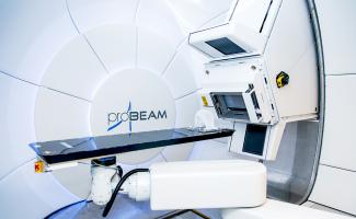
In the world of the robotic radiosurgical system, Cyber-knife is the first modern device designed to treat benign and malignant tumors in any place of the body with unique accuracy. It is a non-invasive treatment method, which is an alternative to the surgical one. It delivers high radiation doses directly to the tumor, significantly reducing the risk of radiation damage to healthy tissues.
The system consists of a compact linear accelerator with an energy of 6 MeV, located on a robotic console, having six degrees of freedom and providing 1200 beam positions during irradiation. The robot manipulator provides movement and guidance of a linear accelerator with high (submillimeter) accuracy.
Treatment on a cyber-knife does not require anaesthesia. The procedure can be performed on an outpatient basis, moreover, it is beneficial for patients who have contraindications for traditional radiotherapy or surgery. To accommodate out-of-town patients, the Center has a day hospital with 50 places.
Preparation for treatment
Treatment with Cyber-knife is preceded by scanning of a tumor or other pathological formation to determine its size, shape and location. Usually scanning begins with a standard CT scan of high resolution (HRCT). To visualize some neoplasms, other methods can also be used such as MRI, angiography or PET diagnostics.
Treatment planning
Images obtained during the scanning process are digitally transferred to the Cyber-knife planning system, where the physician determines the exact size, shape and localization of the lesion. Based on these data, a qualified doctor develops a plan of therapy with the help of special software during which the necessary dose of radiation is also calculated to avoid damage to the healthy tissues surrounding the tumor. At this stage, presence of the patient is not required.
Cyber-knife treatment
During the procedure, the patient in a comfortable position is located on the medical table, which is automatically positioned relative to the Cyber-knife console. Anaesthesia is not required, as the operation on the Cyberknife is painless and non-invasive (does not require injections, incisions and other penetrations into the body). The standard duration of one session (fraction) is 30 to 90 minutes. When irradiated, one or more fractions are used. The cost of treatment, as a rule, is fixed regardless of their number
After treatment
At the end of treatment, patients may experience mild fatigue, which is often associated with relief from an emotional stress due to the successful completion of treatment. The day after radiosurgery, most patients return to their normal everyday life and professional activities.
A detailed interview is conducted with the patient and his relatives about recommendations for follow-up, treatment, lifestyle and medical documentation on the radiosurgery performed is issued. In case of any questions, patients can always contact the doctors of our center by phone or e-mail.
The main method of postoperative monitoring is magnetic resonance imaging, performed with contrast enhancement. With the help of a special program employees of the Radiosurgical Center perform a very accurate overlaying of preoperative and postoperative images to compare them. The scans taken in the MIBS diagnostic centers meet all the requirements necessary for assessing the dynamics. You just need to tell us that this examination is a follow-up procedure after radiosurgical treatment and have an extract about the operation performed available. The received images will be sent via the telemedicine network to St. Petersburg and in three days you will be able to discuss the results of your treatment by phone with the doctors of the radiosurgical center.
If the MRI had not been performed by the MIBS diagnostic centers, then the staffs of the MRI center should be alert about the following requirements for the examination:
- The examination is performed with contrast enhancement.
- The thickness of the slices does not exceed 3 mm.
- The image matrix must be square.
- Pictures should be recorded on CD in DICOM format.
CyberKnife® system is the first and only robotic radiosurgery system in the world designed to treat tumors anywhere in the body with sub-millimeter accuracy.
Clinical evidence
According to statistics, approximately 56% of patients treated with the CyberKnife system in the United States between June 2007 and June 2008, they were cured from tumors located outside the brain. To date a summing up of a total of more than 50,000 patients worldwide. The clinical proof of the CyberKnife system is confirmed by 290 reviews from medical centers.
Main components
X-band compact linear accelerator - generates tumor-damaging radiation beams. Its dimensions and weight are significantly smaller compared to standard medical linear accelerators, which are usually used in radiotherapy. Due to its compact size and low weight, the accelerator can be installed directly on the robotic arm.
The robotic arm - the arm has six degrees of freedom and ensures movement and targeting of the linear accelerator with exceptional accuracy and repeatability from almost any direction. Thanks to this flexibility, a variety of paths and entry and exit points of the beams is achieved, which minimizes the risk of radiation damage to healthy cells near the tumor. Moreover, the short response time of the robotic arm enables tracking in real time the movement of the tumor associated with breathing.
Image guidance system with real-time targeting and feedback - provides continuous tracking, control and synchronization with the tumor and patient movement during the procedure without intervention of a doctor.
Image detectors - capture high-resolution anatomical images during the procedure. These continuously generated images are compared with previously digitized radiographs in order to determine the patient's position in real time. Based on this information, the robotic arm immediately responds to any detected patient movement.
X-ray sources - images are acquired throughout the procedure using low-power X-ray sources to determine the position of reference points formed by the bones. To ensure the highest accuracy of the images, the X-ray sources and image detectors are securely fixed.
Additional technologies
Synchrony® motion synchronization system - constantly synchronizes radiation delivery with the movement of lung, liver or pancreas tumors caused by breathing without the need for holding the breath or synchronizing breathing. Unlike traditional radiotherapy, thanks to the Synchrony system, the patient can breathe normally during the procedure as the system ensures exceptional accuracy and minimal damage to surrounding healthy tissue.
RoboCouch® Patient Positioning System - has six degrees of freedom and performs positioning accurately, quickly and automatically, thereby reducing the duration of the procedure.
Xsight® Spine Tracking System - Using the anatomy of the spinal bones to automatically track the position of the tumor,it eliminates the need for implantation of fiducials. As a result, the need for surgical intervention in the spine is reduced.
Xsight® Lung Tracking System - provides accurate, reliable and direct tracking of the movement of a lung tumor without fiducials.
Xchange ™ Robotic Collimator Changer System – automatically switches collimators, thus increasing the efficiency of conformal procedures.
Four-Dimensional Optimization And Treatment Planning System - tracks not only the movement of the tumor, but also the movement and deformation of surrounding tissues, which optimizes the treatment of tumors being shifted by breathing.
MultiPlan® Treatment Planning System - this intuitive workstation designed specifically for the CyberKnife system allows you to create treatment plans with excellent conformity and high dose gradients.
InView® Image Fusion and Contouring Station – enables remote review of treatment plans and includes necessary tools for ultra-fast image fusion and contouring.
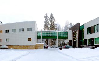
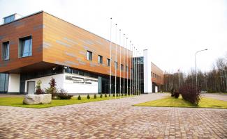
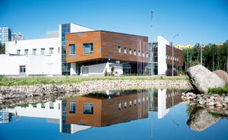
PHOTO AND VIDEOS


Chat with us ![]()
+971581965258
Abu Dhabi, UAE
Abu Dhabi, UAE